A few months ago I rather suddenly developed an acute painful swelling at the base of my left big toe. "I think I'm having an attack of gout, but why me?" I wondered
Of course this had to happen on a weekend, when the Family Practice Clinic I'd usually go to was closed, as was my Podiatrist's office.
I wondered if I could have septic arthritis, an infected joint, but had no obvious reason for this much more frightening diagnosis. I remembered there was also an entity called pseudogout, where the crystals were calcium pyrophosphate, not uric acid.
So I went to the Urgent Care Center our local hospital established a few blocks away from my house. A Family Practice physician examined me and said, "I think you have gout, but you'll need to go to the hospital's Emergency Department (ED) so they can get some fluid from your joint and decide if it's really gout. We don't do that test here." She was also concerned about the rather slim possibility of septic arthritis.
At the ED I was triaged as someone who could wait a while and eventually seen by a Physician's Assistant (PA) who said, "I think you have gout!"
Now I trained at Duke, the home of the original PA program and one of the second generation of PA's taught me how to do dialysis in the ICU for patients with acute kidney failure, so I have no problem at all being seen by a PA.
"What do we do to make sure it's gout and not septic arthritis or pseudogout?" I asked.
"I really don't think you have an infected joint. We'd have to "tap your toe" (i.e., aspirate some fluid from the joint), to make sure it's gout, but that's a tricky procedure, not one that I would do. You should see a podiatrist. In the meantime, take big dose Ibuprofen."
I had a bottle of that at home and knew I could take 800 milligrams three times a day as long as I ate a sandwich or a meal first. I wasn't going to bother the on-call Podiatrist that weekend unless the pills didn't help.
When I did see my regular Podiatrist, two days later, the clinical signs of acute inflammation that I had learned in Latin and English during my first year of medical school:, rubor (redness), calor (increased heat), tumor (swelling), dolor (pain), had all diminished markedly.
"I don't stick joints unless I have to," said my highly experienced Podiatrist. "The PA you saw at the hospital was right; it's not something we do routinely. You're better and this was a classical gout attack, so keep taking the NSAID for a few more days."
When I was in medical school, half a century ago, I thought of gout as a kind of acute arthritis that affected corpulent, older men who ate too much red meat and drank port wine. They then deposited uric acid crystals in joints and sometimes developed a chronic form of the disease called tophaceous gout, where nodular masses of the crystals (tophi) are deposited in different soft tissue areas of the body. Tophi are most commonly found as hard nodules around the fingers, at the tips of the elbows, and around the big toe, but they can appear anywhere in the body, even in the ears, vocal cords, or even against the spinal cord! As a Nephrologist, I was also aware that some people develop uric acid kidney stones.
Here's a link to an article with photos of acute gout in a toe and Henry VIII who suffered from the disease. http://www.dailymail.co.uk/health/article-2210797/Disease-kings-rise-people-gout-increase-obesity.html
I didn't fit the image I had of someone who'd have a gouty attack. I wasn't overweight, didn't drink much (I usually have a glass of wine or whiskey three times a week), didn't have a family history of gout and hadn't over-indulged in the high-purine foods that can increase uric acid levels.
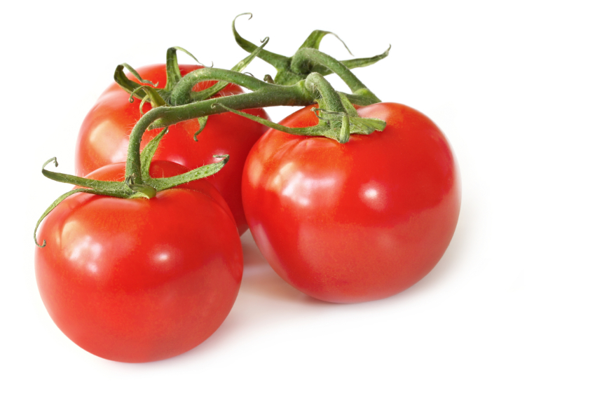
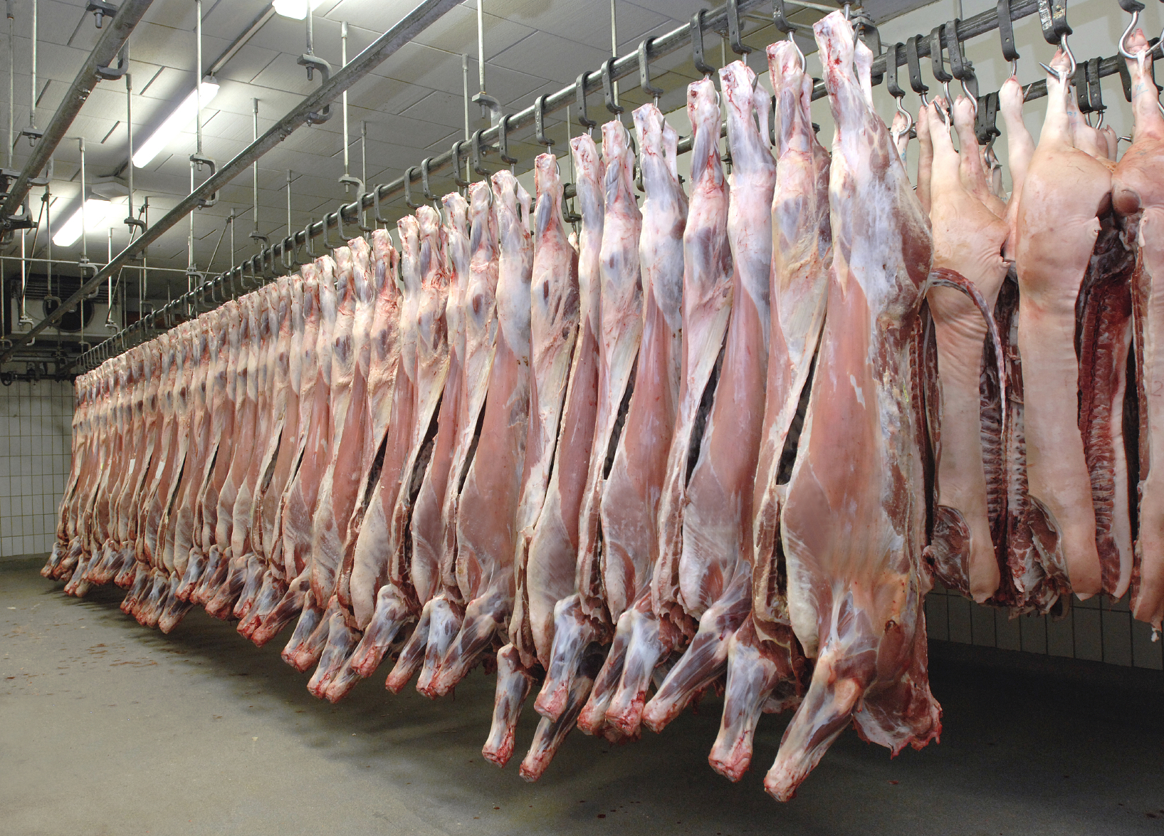
Purines are natural substances found in all of the body's cells, and in virtually all foods. A relatively small number of foods, however, contain concentrated amounts of purines. For the most part, these high-purine foods are also high-protein foods, and they include organ meats like kidney, fish like mackerel, herring, sardines and mussels, and also yeast.
When cells die and get recycled, the purines in their genetic material also get broken down. Uric acid is the chemical formed when purines have been broken down completely. Low-purine diets are often used to help treat severe gout in which excessive uric acid is deposited in the tissues of the body. Purines from meat and fish clearly increase our risk of gout, while purines from vegetables fail to change our risk. Dairy foods (which can contain purines) actually appear to lower our risk of gout.
I had been taking a baby aspirin a day for reasons that were "iffy," and some recent research had implicated even low-dose aspirin as a possible risk factor for gout. Here's a link to that summary online http://www.ncbi.nlm.nih.gov/pubmed/23345599. So I stopped taking aspirin.
I had no personal history of heart disease, although my brother, who had many risks factors, died at 57 of a heart attack and my mother had one at age 74. Aspirin for secondary prevention of cardiovascular events seems to make sense to me (always discuss this with your own physician before you start taking long-term aspirin or any other drug). But primary prevention, that is taking aspirin if you haven't had a heart attack or angina, is a different matter, one that is being hashed over in the medical literature.
I found an article on pseudogout on the WebMD website http://www.webmd.com/osteoarthritis/arthritis-pseudogout and wondered if that's what I had had an episode of. Time would tell.
Then recently I flew to the DC area to visit family and friends, but mostly to hear and see Jordi, my sixteen-year-0ld grandson, who had the male lead roll in "High School Musical," at HB Woodlawn High.
I normally drink three very large glasses of water a day and my family had purchased the limes I squeeze into the water (It tastes better, so I drink more.) But on the flight home I didn't drink much at all and I was aware of pain in my right big toe as I came off the plane.
I woke up at 4:15 a.m. the next morning with fairly severe pain in that toe, took 800 milligrams of Ibuprofen after eating a thick slice of bread abundantly smeared with cream cheese and jam, and went back to sleep.
I was fortunate; my Podiatrist had an appointment cancellation the next morning and he said, "Clinically this is gout. I'm going to give us a "script for another NSAID that I find works better for acute gouty arthritis. And drink lots of fluids"
I went to the pharmacy and got a bottle of indomethacin, an NSAID I haven't used for many years. I had asked him if he wanted me to get a uric acid level blood test and he said, "You can have gout without having an elevated uric acid, but if you do have one, we can treat it. I think the time to get blood tests is after the acute attack resolves.'
I was aware that another old drug used in gout, Allopurinol, was still around, but had to look up its mechanism of action. What I found was Allopurinol reduces the production of uric acid in your body, so if I did have an elevated blood level of uric acid, it could be used to potentially prevent me from having gouty attacks or forming uric acid kidney stones..
I hadn't been aware that an elevated uric acid level might go down during an acute gouty attack; I visualized uric acid crystals migrating to my big toe, but was unsure if that's what the Podiatrist meant.
I'd be very happy if the next episode waits a long time to happen, but I'm not betting on that being the case.